Category | Term | Description |
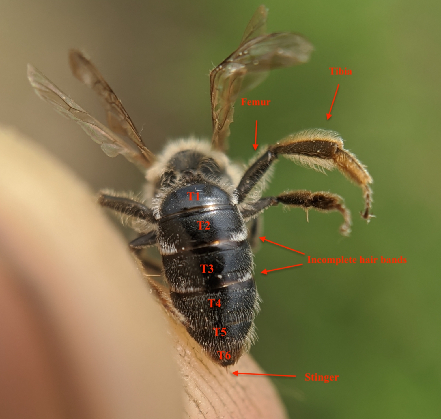
Main (rear) body part | Abdomen | A bee's abdomen is its rear body section, containing vital organs for digestion (honey stomach), reproduction (spermatheca, ovaries, testes), and defense (stinger), plus glands for wax (wax glands) and scent (Nasonov gland) in workers, all covered in protective segments that allow flexibility and house important functions like nectar storage and even breathing through pulsing. T1 is the first segment of the abdomen after the thorax, then T2, T3, T4, T5, and then T6 is the last portion of the abdomen. |
https://val.vtecostudies.org/wp-content/uploads/2021/02/andrena-annotated.jpeg | ||
Main body part (respiration) | Spiracles | Bee spiracles are tiny, valve-controlled holes along a bee's thorax and abdomen that serve as its respiratory openings, allowing air (oxygen) to enter and carbon dioxide to exit, connecting to a network of internal tubes (tracheae) that deliver gases directly to the bee's cells, bypassing lungs. Bees have ten pairs of these spiracles, which open and close to regulate airflow, keeping moisture in and preventing debris from entering, with abdominal movements pumping air in and out. |
Main body part (middle) | Thorax | The thorax is the middle section of a bee's body, and it is the center for locomotion, meaning it's where the six legs and two pairs of wings are attached. This segment contains powerful muscles that drive flight, produce buzzing sounds, and help the bee move. The thorax is also composed of three smaller segments that each have a pair of spiracles (2), which are openings for air. |
Face | Antennae | Sensory appendages for smell, touch, and navigation. |
Face | Antennal Sockets | Cavities in which the antennae articulate. |
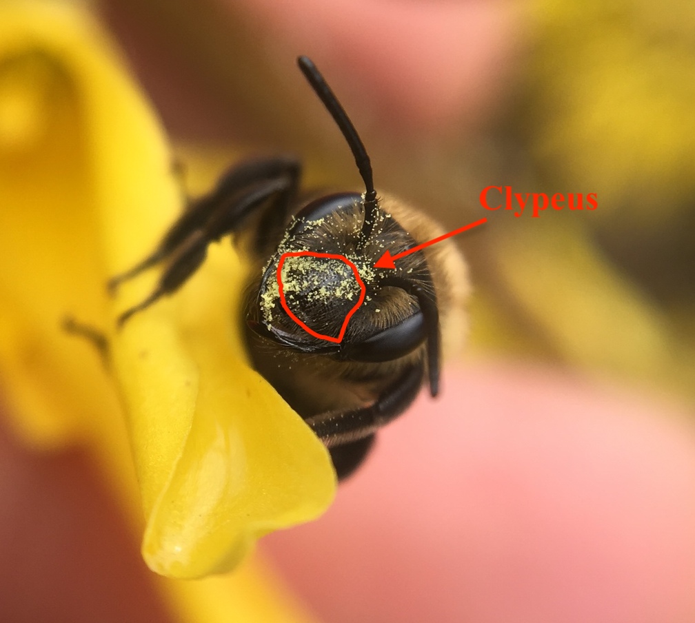
Face | Clypeus | Clypeus refers to the plate on the lower part of an arthropod’s face. Broad facial plate between the frons and labrum. Frons: Central face area between the antennae and ocelli. Labrum: Upper lip assisting food manipulation. |
Face | Compound Eyes | Large, multi-faceted eyes providing wide-angle vision. |
Face detail | https://val.vtecostudies.org/wp-content/uploads/2021/02/clypaeus.jpg | |
Face | Frons | Central face area between the antennae and ocelli. |
Face | Labrum | Upper lip assisting food manipulation. |
Face | Mandibles | Jaws used for cutting, carrying, and nest construction. |
Face | Ocelli | Three simple eyes on top of the head that detect light intensity. |
Face | Paraocular lobe | On a bee's face: an area extending along the sides of the head, parallel to the inner margin of the compound eyes. |
Face | Tooth, or subapical tooth | "Tooth" is a spine-like projection on the bees anatomy, typically located on the mandibles (jaws) or the gena (cheek area behind the eye). These are not true teeth like those found in mammals, but rather hardened, chitinous extensions of the exoskeleton that aid in specific functions. A subapical tooth is a small, tooth-like projection located on the mandible (jaw). |
Mouthparts | Galea | Sheath-like maxillary element forming part of the tongue. |
Mouthparts | Glossa (tongue) | A bee's glossa (Greek for "tongue") is the central, hairy, tube-like structure within its proboscis, acting like a flexible, hairy spoon to lap up nectar, which then gets sucked up the food canal for storage, forming a complex feeding straw. While people often call the whole thing the proboscis, the glossa is just the inner, hairy part that collects liquid, supported by other mouthparts like labial palpi and galeae to create a watertight tube for efficient feeding, making it a "straw-spoon hybrid" |
Mouthparts | Labial Palps | Sensory appendages on the labium. |
Mouthparts | Labium | A bee's labium is its "lower lip," a key part of its sucking mouthparts (proboscis that, along with other structures, forms a tube to drink nectar, acts like a spoon to lap it up, helps clean the bee and hive, and features sensory palps (16) for taste. It's formed from fused ancestral second maxillae and contains the 'tongue' (glossae and paraglossae) and palps for sensory input, allowing bees to taste as they feed. |
Mouthparts | Maxillae | Lateral mouthparts aiding food manipulation. |
Mouthparts | Palps | Bee palps (or palpi) are sensory appendages on a bee's mouthparts, with two pairs—maxillary palps (upper) and labial palps (lower)—that help with sensing food, grooming, and manipulating the proboscis (tongue) for nectar feeding. These jointed feelers detect chemicals and textures, aiding in food selection and taste, acting like tiny sensory antennae near the mouth. |
Mouthparts | Paraglossa | Small lobes flanking the main glossa. Glossa: Tongue |
Mouthparts | Proboscis / Glossa | Elongate tongue used for nectar uptake. |
Dorsal | Mesoscutum | Large central dorsal (top) thorax plate. |
Dorsal | Metanotum | Small plate behind the scutellum. Scutellum: Plate following the mesoscutum. |
Dorsal | Pronotum | Narrow segment behind the head. |
Dorsal | Propodeum | First abdominal segment fused to thorax. |
Dorsal | Sclerite | A sclerite is any of the hardened plates that form the bee's exoskeleton. These rigid, chitinous plates are connected by flexible, soft membranes (arthrodial membranes), which allow the bee to move despite having a hard outer covering. |
Dorsal | Scutellum | Plate following the mesoscutum. (Small section at the base of the Thorax). Mesoscutum: Large central dorsal (top) thorax plate. |
Dorsal | Tegulae | Small sclerites above wing bases. (Where the wing attaches to the thorax.) |
Dorsal | Tergites | Dorsal (upper or back of the bee) abdominal segments. |
Dorsal | Vertex | Top of the head behind the ocelli. Ocelli: Three simple eyes on top of the head that detect light intensity. |
Ventral | Corbicula Floor | Surface supporting pollen loads. |
Ventral | Gena | Lower side portion of the head. |
Ventral | Mesepisternum | Major thoracic side plate. |
Ventral | Sternites | Ventral (underside) abdominal segments. |
Ventral | Ventral Propodeal Plate | Underside of the propodeum. Propodeum: First abdominal segment fused to the thorax. |
Lateral | Gena | Cheek region behind the eyes. |
Lateral | Mesepisternum | Large thoracic plate on the side. |
Lateral | Metepisternum | Rear side plate of thorax. |
Lateral | Pre-episternal Groove | Groove separating thoracic plates. |
Lateral | Propodeal Spiracle | Respiratory opening on propodeum. Propodeum: First abdominal segment fused to the thorax. |
Lateral | Temporal Area | Posterior-lateral head area. P-L: rear and on the side of the head. |
Leg | Arolia | Adhesive pads for traction. |
Leg | Corbicula | Pollen basket on hind tibia. Corbicula (Corbiculae pl.) refers to the “pollen basket”, a part of the hind legs of some bee species such as bumble bees and honey bees. Corbiculae are used to harvest and carry pollen from flowers to the nest or hive. Other bees have scopae. Corbiculae - Many bees have flattened plates used as pollen baskets on hind legs (e.g. honey bees (Apis mellifera) and bumble bees (Bombus spp.). Tibia: Segment bearing spines and scopa. |
Leg | Coxa | Basal leg segment attaching to thorax. |
Leg | Femur | Robust upper leg segment. |
Leg | Scopa | Pollen-collecting hairs. Scopa (Scopae pl.) refers to the groups of hairs on the body of some bees that help in pollen collection and transport from flowers to the nest. Scopae - Pollen-carrying hairs usually on the hind legs or the underside of the abdomen, often covered with pollen. |
Leg | Tarsal Claws | Terminal claws for gripping. |
Leg | Tarsus | Multi-segmented foot. |
Leg | Tibia | Segment bearing spines and scopa. The Tibia follows the femur. Scopa: Pollen-collecting hairs. |
Leg | Trochanter | A small segment between the coxa and femur. Coxa: Basal leg segment attaching to the thorax. Femur: Robust upper leg segment. |
Wings | Costa | Leading wing edge vein. |
Wings | Forewing | Larger anterior wing. Anterior: near the front. |
Wings | Hamuli | Hooks connecting the hindwing to the forewing. Back and front wings. |
Wings | Hindwing | Smaller wing attaching via hamuli. Hamuli: Hooks connecting the hindwing to the forewing. Back and front wings. |
Wings | Radius/Media/Cubitus | Primary longitudinal veins. (Long.: running lengthwise) |
Wings | Stigma | Thickened leading-edge area. |
Wings | Subcosta | Second major vein behind the costa. Costa: Leading wing edge vein. |
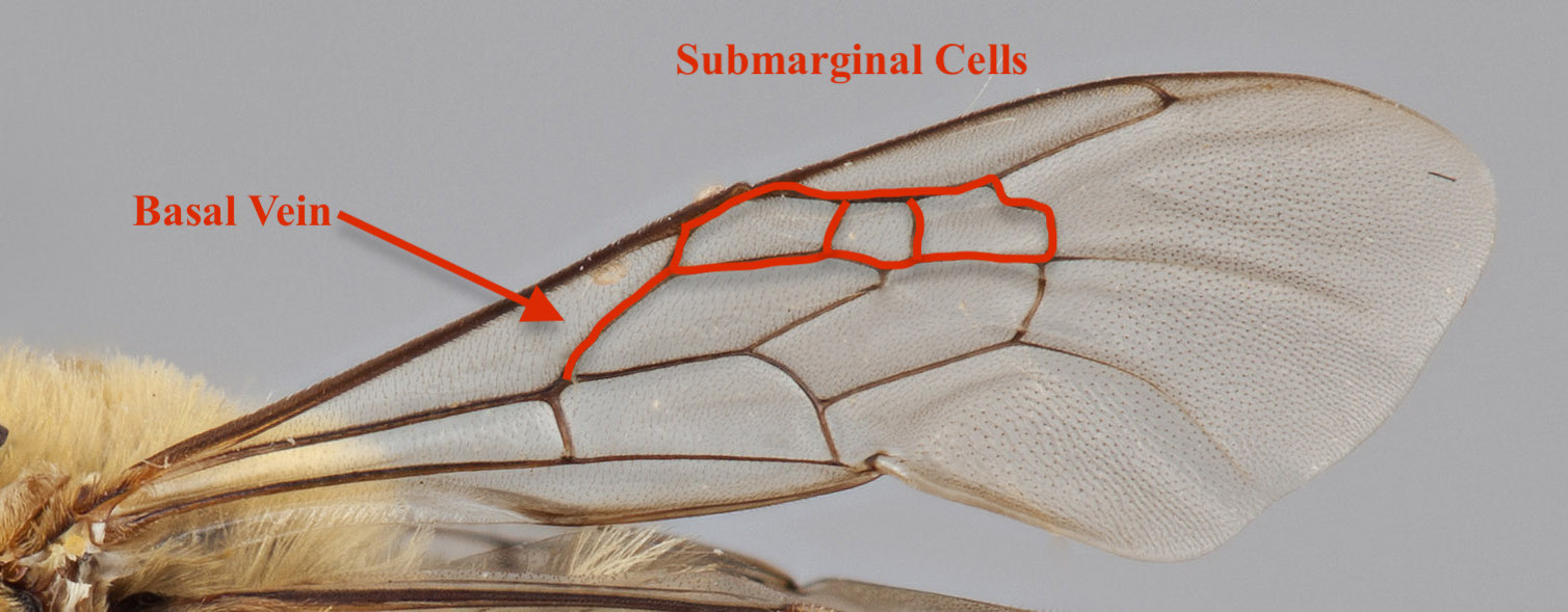
Wings | Wing Cells | Enclosed wing areas are used in taxonomy. |
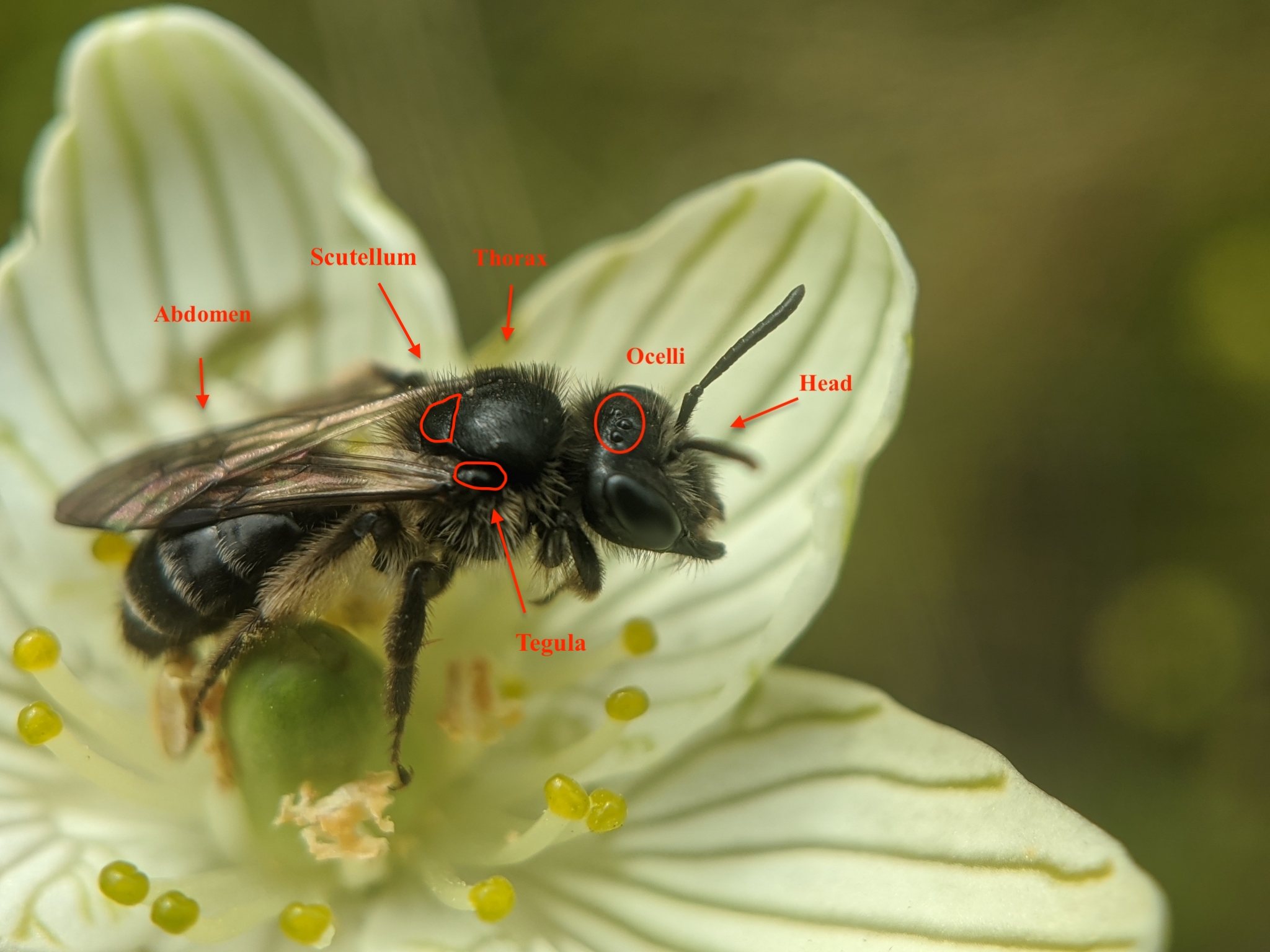
Important morphology (VCE) | General Body plan of a bee (Photo) | https://val.vtecostudies.org/wp-content/uploads/2021/02/body-parts-2.jpeg |
Full Anatomy | Labelled diagrams | |
Wing detail | https://val.vtecostudies.org/wp-content/uploads/2021/02/Wing.jpg |
Link to: Bee Anatomy titled Illustrations
No comments:
Post a Comment
Thank you for your comment. Your comment will appear once the moderator views and accepts it.